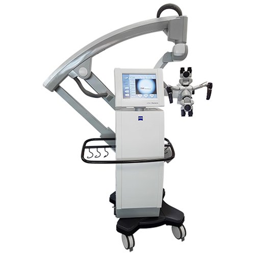
Zeiss OPMI Pentero
The Zeiss OPMI Pentero surgical microscope features apochromatic optics that deliver crystal-clear images, sharp details, and natural colors. The OPMI Pentero has 20% more light than previous models with spot illumination to precisely adjust the light cone. The Pentero has integrated high-speed autofocus that automatically delivers sharp images regardless of magnification. With the overhead design of this microscope, the suspensions system can be placed in any position, even behind the surgeon.
- Automated functions such as AutoBalance and AutoDrape
- Image-guided surgery with MultiVision™ data injection
- Integrated digital visualization, optionally with integrated high-definition (HD) camera head
- DICOM networking capabilities
- Touchscreen operation
Zeiss OPMI Pentero Specifications
Dimensions
- Height: 81.1” (206 cm)
- Width: 28.97” (73.6 cm)
- Depth: 28.97” (73.6 cm)
Magnification System
- Motorized zoom, apochromatic, 1:6 ratio
- Magnification displayed on the touchscreen and in the ocular (on demand)
- User-specific start position
Focusing System
- Varioskop, apochromatic, 200–500 mm working range
- Internal, motorized, continuous adjustment
- Magnification linked adjustment of focus speed
- High-speed laser autofocus, accurate to +/- 0.5 mm (Class II Laser)
- Visual focusing aid with two converging laser spots
- Working distance displayed on a touchscreen and in the ocular (on demand)
- User-specific start position
MultiVision System
- Integrated data display with shutter function
- SVGA 800 x 600, color, 50-60 Hz
- Color, binocular, injection and superimposition of contours and data
- Supported external data signals
- Computer data (VGA Signal)
- I.e. data from navigation systems
- Computer data (VGA Signal)
-
- Y/C video data (PAL / NTSC)
- I.e. data from endoscopy systems
- Y/C video data (PAL / NTSC)
- Superimposition of system information (focus, zoom, light)
- Injection of the touchscreen user interface into the eyepiece for sterile control of the system
Tubes and Co-Observation
- Main tube: 0–180° rotatable
- Eyepieces 10x/21B, 12.5x/18B
- Integrated beam splitter for lateral and face-to-face co-observation
- Stereo co-observation tube remains fixed when tilting the OPMI
- Spine adapter for symmetric face-to-face configurations
- Integrated rotary tube adapters
AutoDrape Systems
- Integrated vacuum system to remove air from sterile drape for fast and easy draping
Illumination System
- Superlux 330 light source with two 300 W Xenon daylight character lamps
- Integrated light source and light guide
- Integrated two-way illumination brightens shadows
- Variable spot illumination, minimum diameter 10 mm
- Semi-automatic lamp exchange
- Display of remaining lamp life on Touchscreen
- Brightness regulation via handgrips
- Magnification dependant automatic brightness adjustment
- Synchronized camera flash system
AutoBalance
- AutoBalance of the microscope, suspension system or entire system by pushing a button
- Microscope AutoBalance independent of position or accessories
Hospital Workflow Integration
- Varioskop, apochromatic, 200–500 mm working range
- Internal, motorized, continuous adjustment
- Magnification linked adjustment of focus speed
- High-speed laser autofocus, accurate to +/- 0.5 mm (Class II Laser)
- Visual focusing aid with two converging laser spots
- Working distance displayed on a touchscreen and in the ocular (on demand)
- User-specific start position
MultiVision System
- LAN interface and modem
- Microphone and speaker
- Patient data management allowing archival of image, video and audio data service file
- Remote service interface
Integrated Digital Video Chain
- 3CCD-Video camera PAL/NTSC
- Video output on touchscreen
- Digital video outputs: Firewire/DV and Progressive Scan (VGA)
- Analog video outputs: FBAS (BNC), Y/C, RGB
- Stereo camera
- Image capture
- Image freeze function
- Image capture as TIFF, JPG, BMP
- Image annotation
- Still image archiving via CD/DVD/USB and optional DICOM** interface
- Digital video recording system:
- MPEG2 recording
- Parallel HD/DVD recording
- Editing function
