SonoSite M-Turbo Ultrasound Machine
January 4, 2018
SonoSite M-Turbo Ultrasound Machine
What is an Ultrasound Machine?
Ultrasound imaging uses sound waves to produce pictures inside the body. It is used to help diagnose the causes of pain, swelling, and infection in the body. Most people know them to examine a fetus in a pregnant woman or the brain and hips in infants. They can also be used to help guide biopsies, diagnose heart conditions, and assess damage after a heart attack. Ultrasounds are very safe and noninvasive. They do not use ionizing radiation.
Ultrasound imaging is also called ultrasound scanning or sonography. It involves the use of a small transducer or probe and ultrasound gel that is placed directly on the skin. High-frequency sound waves are transmitted from the probe through the gel and into the body. The transducer collects the sounds that bounce back and a computer uses them to create an image. Ultrasound images are captured in real-time, they can show the structure and movement of the body’s internal organs, as well as blood flowing through blood vessels.
Common Ultrasound Uses of Ultrasound Machines
Ultrasound examinations can help diagnose a variety of conditions and to assess organ damage following an illness. It can be used to help diagnose the following symptoms.
- Pain
- Swelling
- Infection
What parts of the body can be examined?
Ultrasound is a useful way of examining many of the body’s internal organs, including but not limited to:
- The Brain, Spine, and hips in infants
- Scrotum
- Thyroid and parathyroid glands
- Eyes
- Uterus, ovaries, and a fetus in pregnant patients
- Bladder
- Kidneys
- Pancreas
- Spleen
- Gallbladder
- Liver
- Heart and blood vessels
SonoSite M-Turbo Features
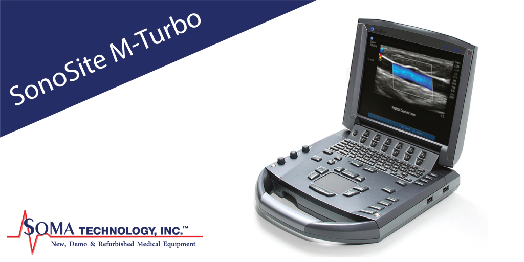
The SonoSite M-Turbo portable ultrasound is SonoSite’s most versatile system for abdominal, never, vascular, cardiac, venous access, pelvic, and superficial imaging. The M-Turbo ultrasound machine comes with SonoGT and SonoHD imaging technology, enhanced color flow, optional SonoRemote control. The M-Turbo has 16 times the processing power of the previous SonoSite generation and still weighs under 7 pounds.
SonoGT
SonoGT Global Targeted technology is an advance that capitalizes on the power of the M-Turbo platform to dive a new level of color flow imaging, wireless connectivity and workflow integration for anesthesia, emergency medicine, critical care, and other acute point-of-care markets.
ColorHD
ColorHD Technology is a proprietary, color Doppler algorithm that is available on all transducers. This technology works in parallel with multiple M-Turbo algorithms, including SonoGT to provide increased diagnostic information and better visualization of color flow, specifically in low flow states.
SonoRoam
Using 802.11b/g technology, SonoRoam enables wireless image transfer from the M-Turbo system to an existing PACS system via DICOM or to a personal computer via SonoSite SiteLink so clinicians can quickly retrieve the information from any location.
SonoSite M-Turbo Specifications
Dimensions
- Weight: 6.7 lbs (3.04 kg) (without battery and transducer)
- Dimensions: 11.9” L x 10.8” W x 3.1” H (30.2 cm L x 27.4 cm W x 7.9 cm H)
- Display: 10.4” (26.4 cm) diagonal LCD (NTSC or PAL)
Imaging Modes and Processing
- Broadband, multi-frequency imaging.
- 2D/Tissue Harmonic Imaging/M-Mode.
- Velocity Color Doppler.
- Color Power Doppler.
- PW, PW Tissue Doppler, and CW.
- Doppler angle, correct after freeze.
Image processing
- SonoADAPT™ Tissue Optimization.
- SonoHD™ Imaging.
- SonoMB® Multi-beam Imaging.
- Advanced Needle Visualization.
- Auto gain automatic image optimization.
- Dual Imaging.
- Duplex Imagine.
- 2x pan/zoom capability.
Transducers
- Broadband and multifrequency.
- Linear Array, Curved Array, Phased Array, Multiplane TEE, and Micro-Convex.
Single frequency
- Cardiac Static Pencil.
USB Storage Formats
- MPEG-4 (H.264), JPEG, BMP, HTML.
- Compatible with Mac® and PC formats.
User Interface and Remappable Controls
- Softkeys to drive advanced features.
- Programmable A and B keys: each can be assigned by the user for increased ease of use.
- Alphanumeric elastomeric QWERTY keyboard.
- Trackpad with the select key for easy operation and navigation.
- Doppler controls: angle, steer, scale, baseline, gain and volume
- Image acquisition keys: review, report, Clip Store, DVD, save.
- Dedicated AutoGain and exam keys to allow quick activation.
Application-Specific Calculations
- Cardiac: Complete cardiac calculations package and patient report, including ventricular, aortic and atrial measurements, ejection fraction, volume, Simpson’s rule, continuity equation, pressure half-time, and cardiac output.
- OB/GYN/Fertility: Diameter/ellipse measurements, volume, six follicle measurements, estimated fetal weight, established due date, gestational age, last menstrual period, growth charts, user-defined tables, multiple user-selectable authors, ratios, amniotic fluid index, patient report.
- Vascular: Diameter/ellipse/trace measurements, volume, volume flow, percent diameter and area reduction, Lt/Rt CCA, ICA, ECA, ICA/CCA ratio, time average mean (TAM), peak trace, ICA/CCA ratio, angle correction, patient report.
- IMT (Intima Media Thickness): Embedded SonoCalc® IMT software – automatic edge detection with mean and maximum thickness reporting.
Onboard Image and Clip Storage/Review
- 8 GB internal Flash memory storage capability.
- Potential to store 30,000 images or 960 2-second clips.
- Clip Store capability. (maximum single clip length: 60 seconds.)
- Clip Store capability via either the number of heart cycles (using the ECG) or time base.
- Maximum storage in ECG beats mode is 10 heart cycles.
- Maximum storage in time base mode is 60 seconds.
- Cine review up to 255 frame-by-frame images.
Power Supply
- System operates via battery or AC power
- Rechargeable lithium-ion battery.
- AC: universal power adapter, 100-240 VAC, 50/60 Hz input, 15 VDC output.
Measurement Tools, Pictograms, and Annotations
- 2D: Distance calipers, ellipses, and manual trace.
- Doppler: Velocity measurements, pressure half-time, auto and manual trace.
- M-Mode: Distance and time measurements, heart rate calculation.
- User-selectable text and pictograms.
- User-defined, application-specific annotations.
- Biopsy guidelines.
- External Data Management and Wireless
- DICOM® Image Management. (TCP/IP)
- Print and Store.
- Modality Work List.
- Storage Commit
- Modality Performed Procedure Step.
- SiteLink™ Image Manager functionality.
- SonoSite® Education Key™ training video compatible.
- SonoSite Workflow Solutions.
- SonoRemote™ Control – Bluetooth® wireless technology.
External Video and Audio
- S-video (in/out) to VCR or DVD for record and playback.
- RGB or DVI output to external LCD display
- Composite video output (NTSC/PAL) to VCR or DVD, video printer or external LCD display.
- Audio output.
- Integrated speakers.
Supported Peripheral Devices
- B/W video printer.
- DVD recorder.
- barcode reader
Explore Other Blog Items By Category
Recent Posts

May 29, 2026
May 27, 2026
How do Bladder Scanners Measure Urine?
April 15, 2026